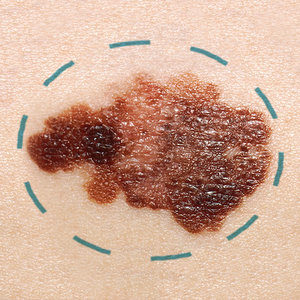
We want to help keep your skin healthy and beautiful from head to toe. Sometimes it is medically necessary to remove portions of your skin surgically to ensure your overall health.
River Ridge dermatologists are board-certified and are dedicated to your health and the health of your skin.
We only perform medically necessary procedures or desired cosmetic procedures to maintain the health, function, and appearance of your skin.
Advancements in surgical techniques allow your dermatologist to ensure that procedures are effective and beautiful. More than ever before, dermatologic surgical procedures are less intense, safer, and heal more quickly.
Some dermatological surgery procedures we provide include excisions, flaps, grafts, nail surgery, laser surgery, and chemical peels.
Below are a few forms that explain what you need to do to take care of your skin after a surgical procedure. If you have a surgery scheduled, or are considering a surgical option, please read the attached documents and give us a call if you have any questions or concerns.
MOHS SURGERY

Mohs surgery is a precise surgical technique that targets cancerous skin cells while minimizing damage to surrounding healthy tissue.
Mohs surgery is done in short stages with lab work processed on-site while you wait.
Mohs surgery — the steps
Step 1. A surgeon trained in Mohs surgery, pathology, and reconstructive surgery examines the cancerous spot, possibly drawing some marks on the patient’s skin for reference, then injects a local anesthetic to numb the area.
Step 2. Using a scalpel, the surgeon removes a thin layer of visible cancerous tissue.
Step 3. While the patient’s wounds are being temporarily bandaged, an on-site technician processes the removed tissue. The surgeon analyzes it to determine if the cancer extends deeper beneath the skin. If it does, the patient will be called back into the operating room to have another layer of tissue removed.
Step 4. The team repeats the whole process until the edges of the last excised tissue sample are free of visible cancer.
Step 5. If needed, the surgeon closes the wound with stitches or a skin graft.
Step 6. The surgery is complete. The patient is given instructions on caring for the incision site.
POST-SURGICAL CARE:
Below are a few forms that explain how to take care of your skin after a surgical procedure. If you have a surgery scheduled, or are considering a surgical option, please read the attached documents. Call us if you have any further questions or concerns.
Post-surgical care information sheets:
What it is:
Basal cell carcinoma, or BCC, is an abnormal, uncontrolled growth or lesion that arises in your skin’s basal cells—the deepest layer of the epidermis. BCC is a non-melanoma skin cancer.
BCCs look like open sores, red patches, pink growths, shiny bumps, or scars. They are usually caused by sun exposure.
As long as they are treated, BCC does not usually spread, but are serious and can be disfiguring if not removed quickly. Having regular skin checks is key to early detection.
Treatment:
BCCs are diagnosed with a biopsy. Depending on how the BCC presents and the needs of the patient, the BCC can be removed surgically, treated with radiation, cryosurgery (freezing), or photodynamic therapy; or topical or oral medications.
What they are:
A benign lesion is non-cancerous. It will not spread abnormal cells, and should not threaten your health. However, these lesions may still need to be surgically removed due to location, size, and appearance.
A malignant lesion, such as basal cell carcinoma, squamous cell carcinoma, or melanoma, has been biopsied and determined cancerous. It is characterized by progressive and uncontrolled growth. This kind of lesion is dangerous and needs to be surgically removed.
Treatment:
How a lesion is removed depends on the type of lesion and where it is located.
Surgical excision:
This is a simple form of plastic surgery. The lesion and area around it are numbed with an anesthetic, and the doctor makes an incision through the full three layers of your skin—the epidermis, dermis, and the subcutaneous fat.
The doctor removes the specimen, then pulls the edges of the wound together using plastic surgery techniques. This provides a better cosmetic end result (as opposed to methods using scraping or burning). The cure rate for this technique is very high.
Electrodessication and Curettage (Scraping and burning):
For basal cell carcinomas that are superficial and confined to the epidermis, or top layer of the skin, one of the most effective treatments is electrodessication and curettage, which is a technique involving scraping and burning. This procedure is relatively quick and easy, but can only be used on basal cell lesions on the arms, legs, and trunk. It does leave a small, pale scar.
What they are:
Cysts are benign skin lesions that generally do not require treatment. However, they have the potential to enlarge and become inflamed in which case patients may elect to have them surgically removed.
What it is:
A non-cancerous growth of fatty tissue cells that can develop in almost any organ of the body, although they are most commonly found just below the skin. Lipomas are very common, grow slowly and are not painful. Generally, lipomas do not require treatment; however, they can be removed with a small skin incision under local anesthesia.
Treatment:
Excision:
The doctor may cut out the lipoma in rare situations.
What it is:
Melanoma is the most dangerous form of skin cancer. These cancerous growths develop when unrepaired DNA damage to skin cells, caused by UV radiation from the sun or tanning beds, triggers mutations. The skin cells then multiply rapidly and form malignant tumors.
Melanomas often resemble moles. Some develop from moles. Most melanomas are black or brown, but they can also be skin-colored, pink, red, purple, blue, or white.
If melanoma is detected and treated early, the cure rate is excellent. If a melanoma is not caught in time, the cancer can spread to other areas of the body and be deadly. Not all melanomas are caused by sun exposure. It is important to look everywhere, even in areas that are not exposed.
Use the ABCDE’s to evaluate your skin:
A-Asymmetry, meaning one half doesn’t look like the other
B-border irregularity, a jagged or scalloped edge
C-color, either multiple colors in one lesion or one very dark color
D-diameter, anything greater than 6mm or the size of a pencil eraser
E-evolution, some sort of change
Treatment:
The first step can be to remove the melanoma surgically. Surgical techniques have improved in the last decade, and much less tissue is removed during the excision, scars are smaller, and the procedure has a faster recovery and is easier to tolerate. Mohs micrographic surgery may also be a good option to remove your melanoma.
In most cases, the surgery can be done as an outpatient procedure under local anesthetic or in the doctor’s office.
If your cancerous melanoma has advanced to stage 3 or 4, you may need additional therapies, such as chemotherapy, immunotherapy, or other targeted therapies, to ensure the cancer is eradicated.
Everyone needs to have regular skin checks with the doctor for early detection and to make sure that no further melanomas develop.
What it is:
Skin cancer is an abnormal growth of skin cells and can be divided into two categories: melanoma and non-melanoma. Skin cancer most often develops on the areas of the skin that are exposed to the sun. Skin cancer affects people of all colors and races. Regular and thorough application of sunscreen with a minimum SPF 30 is the most effective method of preventing the development of skin cancer.
Skin cancer can manifest in a variety of ways: atypical moles, actinic keratosis, basal cell carcinoma, squamous cell carcinoma, and melanoma.
If caught early, skin cancer can be treated relatively easily. However, skin cancer can be dangerous, and in some cases, fatal.
Use the ABCDE’s to evaluate your skin:
A-asymmetry, meaning one half doesn’t look like the other
B-border irregularity, a jagged or scalloped edge
C-color, either multiple colors in one lesion or one very dark color
D-diameter, anything greater than 6mm or the size of a pencil eraser
E-evolution, some sort of change
Watch for anything suspicious including lesions that are new, grow or change rapidly, bleed easily, or do not heal. Keep an eye out for any new or changing moles. If you have a mole or skin lesion that looks suspicious, call us to make an appointment today.
Treatment:
Every occurrence of skin cancer is different, and may require different kinds of treatments. Depending on the type, skin cancer can be treated by various methods including:
Surgery:
Surgery options for skin cancer lesions include
- Excision: the tumor is cut from the skin, along with some of the normal skin around it.
- Shave excision: the abnormal area is shaved off the surface of the skin with a small blade.
- Curettage and electrodesiccation: the tumor is cut form the skin with a curette (a sharp, spoon-shaped tool). A needle-shaped electrode is used to treat the area with an electric current to stop the bleeding and destroy the remaining cancer cells at the edge of the wound. The process may be repeated one to three times during the surgery to ensure all of the cancer is removed.
- Cryosurgery: a treatment that freezes and destroys abnormal tissue.
Laser surgery: a procedure that uses a narrow beam of intense light like a knife to make bloodless cuts in tissue or to remove a surface lesion, like a tumor. - Dermabrasion: removal of the top layer of skin using a rotating wheel or small particles to rub away skin cells.
- Radiation: a cancer treatment using high energy x-rays and other types of radiation to kill cancer cells, or at least prevent them from growing. External radiation therapy uses a machine outside the body to send radiation toward the cancer. Internal radiation therapy uses a radioactive substance and is placed directly into or near the cancer. The way radiation therapy is given depends on the type and stage of cancer being treated.
Chemotherapy:
A cancer treatment using drugs to stop the growth of cancer cells. It can be taken orally or injected into a vein or muscle. The drugs enter the bloodstream and reach cancer cells throughout the body (systemic chemotherapy).
Chemotherapy for non-melanoma skin cancers and actinic keratosis is usually topical (applied to the skin as a cream or lotion). The way chemotherapy is given depends on the type and stage of cancer being treated.
Photodynamic therapy:
Uses a drug and blue light to kill cancer cells. The drug is not active until it is exposed to light and injected into the patient. This therapy causes little damage to healthy tissue.
Biologic therapy:
Uses your own immune system to fight cancer. Substances made by the body or in a lab can boost, direct, or restore the body’s natural defenses against cancer. This kind of treatment is also called biotherapy or immunotherapy. Interferon and imiquidmod are frequently used to treat skin cancer.
Clinical trials:
Taking part in a clinical trial may be a good treatment choice for some patients. Clinical trials are performed to test new treatments to see if they are safe and effective. Talk to your doctor if you are willing to take part in a clinical trial.
What it is:
Squamous cell carcinomas are non-melanoma skin cancers and are usually caused by sun exposure. It is the second most common skin cancer, after basal cell carcinoma. Many squamous cell skin lesions are on the head and neck, and they can look like open sores, red patches, or have a wart-like appearance. It is not often fatal, but surgery for advanced-stage disease can be disfiguring.
Treatment:
A low-risk squamous cell lesion that is not on the face is usually treated with electrodessication and curettage. For an invasive squamous cell carcinoma, a surgical excision or Mohs micrographic surgery can be the best treatment options. Radiation is frequently used after a surgical procedure to ensure that all cancer cells are destroyed. In high-risk cases, chemotherapy may also be used after surgery.
Having regular skin checks is key to early detection and management. It is critical for patients who have had squamous cell lesions to avoid exposure to UV rays and always wear sunscreen.
What they are:
Warts are benign growths caused by the human papillomavirus. They can present anywhere on the skin, but the hands, feet, knees, face, and genitals are the most common. There are more than 100 different kinds of warts.
Treatment:
Warts are very difficult to treat. No one treatment is uniformly effective, so your doctor may suggest the least expensive, easiest treatment at first. Multiple treatments are usually necessary.
There is no cure for the wart virus, so this means that warts can return at the same site or appear in a new spot.
Some treatments include:
Benign neglect:
65 percent of warts disappear spontaneously within two years. Although without treatment, patients risk warts that may enlarge or spread.
Topical agents:
Salicylic acid, available over-the-counter, can be 70-80% effective.
Cantharidin, dibutyl squaric acid, trichloroaceitc acid, podophyllin, or amniolevulinic acid, are administered by trained personnel in the doctor’s office. Or prescription medications like imiquimod or cidofovir, can be applied by patients at home.
Intralesional injections:
Persistent warts that don’t respond to topical agents may require intralesional injections. Candida, Trichophyton, Belomycin, and Interferon-alfa have all be used with varying degrees of success.
Photodynamic therapy:
Certain warts respond to photodynamic therapy, which involves the patient being injected with photoreactive chemicals. The patient is then exposed to light strong enough to activate the chemicals, which causes them to destroy the targeted abnormal cells. This treatment can also be used to treat basal and squamous cell carcinomas, actinic keratosis, psoriasis, acne, and wrinkle rejuvenation.
Systemic agents:
Systemic medications are prescription drugs that work throughout the body. Systemic medications are used when other treatments are not responsive, or the patient cannot take topical medications or UV light therapy. Systemic agents are also used to treat severe psoriasis, psoriatic arthritis, acne, and other dermatological diseases.
Cryotherapy:
For common warts in adults and older children, cryotherapy, or freezing, is a common treatment. It is not painful, but may cause dark spots in people who have dark skin. Sometimes this procedure needs to be repeated more than once to be completely effective.
Electrosurgery and curettage:
Electrosurgery, or burning, is a good treatment for common warts, filiform warts, and foot warts. Curettage involves scraping off the wart with a sharp knife or small, spoon-shaped tool. These two procedures are often used together. The dermatologist may remove the wart by scraping it off before or after electrosurgery.
Excision:
The doctor may cut out the wart in rare situations.
